1st Edition
CTscan and post-processing for life and geosciences
December 2nd-6th, 2019, Oslo (Norway)


Imaging
REGISTRATION IS CLOSED
Please, SUBSCRIBE if you want to receive information on new editions

The course gives a practical introduction to microfocus X-ray tomography (CT scanning) and post-processing of volume data, as used in zoology, botany, paleontology, geology and archaeology.
A Nikon Metrology XT H 225 ST instrument at the Natural History Museum in Oslo will be available to participants throughout the course. This flexible instrument can scan objects from about 2 mm to 20 cm across, with densities ranging from soft tissues to rocks, allowing demonstration of a range of applications and scanning parameters.
Visualization and post-processing of data will be based on the advanced software Avizo, available at most major research institutions.
We will also give a short overview to data analysis methods such as volumetric and texture analysis and geometric morphometrics.
Course instructor will be Dr. Øyvind Hammer at the Natural History Museum, University of Oslo. He has worked with microfocus X-ray tomography for many years and has produced many thousand scans for a wide variety of scientific applications. He has a background in computer science, physics and palaeontology. Dr Anna Nele Herdina will cover the use of contrasting agents (DiceCT).
All participants must bring their own personal laptop.
Natural History Museum
Premises at Økern
Kabelgaten 40, Oslo, Norway
December 2nd-6th, 2019
English
30 hours on-site.
This course is equivalent to 1 ECTS (European Credit Transfer System) at the Life Science Zurich Graduate School.
The recognition of ECTS by other institutions depends on each university or school.
Places are limited to 15 participants and will be occupied by strict registration order. If the course fills up there will be an assistant instructor to help during the practise time.
Participants who have completed the course will receive a certificate at the end of it.

Dr. Øyvind Hammer
University of Oslo
NorwayUniversity of Exeter
United Kingdom

Dr. Anna Nele Herdina
Medical University of Vienna
Austria

Dr. Soledad De Esteban-Trivigno
Transmitting Science
Spain

Dr. Hugo de Boer
University of Oslo
Norway
Contact: courses@transmittingscience.org
Daily Program
Monday
Tuesday
Wednesday
Thursday
Friday
You can check the list of Ambassador Institutions. If you want your institution to become a Transmitting Science Ambassador please contact us at communication@transmittingscience.com
The class schedule is approximate; it is possible that the content of one day may run into the next and a working day may be longer than advertised.
Discounts are not cumulative and apply only on the Course Fee. We offer the possibility of paying in two instalments (contact courses.crete@transmittingscience.org).
5 % Discount
Former participants will have a 5 % discount on the Course Fee.
20 % Discount
20 % discount on the Course Fee is offered for members of some organizations (Ambassador Institutions). If you want to apply to this discount please indicate it in the Registration form (proof will be asked later).
40 % Discount
Unemployed scientists living in Spain, as well as PhD students based in Greece without any grant or scholarship to develop their PhD, could benefit from a 40 % discount on the Course Fee. If you want to ask for this discount, please contact the course coordinator. That would apply for a maximum of 2 places and they will be covered by strict inscription order.


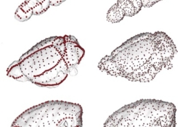 Gonzalez et al. 2016. American Journal of Physical Anthropology